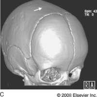
Surgeons and Georgia Tech engineers are working together to improve the treatment of babies born with craniosynostosis, a condition that causes the bone plates in the skull to fuse too soon. Treating this condition typically requires surgery after birth to remove portions of the fused skull bones, and in some cases the bones grow together again too quickly -- requiring additional surgeries.
Researchers in the Atlanta-based Center for Pediatric Healthcare Technology Innovation are developing imaging techniques designed to predict whether a child’s skull bones are likely to grow back together too quickly after surgery. They are also developing technologies that may delay a repeat of the premature fusion process.
“Babies are usually only a few months old during the first operation, which lasts more than three hours and requires a unit of blood and a stay in the intensive care unit, so we are ultimately trying to develop technologies that will simplify the initial surgery and limit affected babies to this one operation,” said center co-director Joseph Williams, clinical director of craniofacial plastic surgery at Children’s Healthcare of Atlanta at Scottish Rite and clinical assistant professor in the Department of Plastic and Reconstructive Surgery at Emory University
“Following the first surgery, there’s a clinical need to be able to screen children on a regular basis to predict when their skull bones are going to fuse together again so that the surgeons can determine if additional intervention will be required,” said center director Barbara Boyan, the Price Gilbert, Jr. Chair in Tissue Engineering in the Wallace H. Coulter Department of Biomedical Engineering at Georgia Tech and Emory University.
To address this need, the researchers have developed a non-invasive technique to monitor bone growth with computed tomography images. They created software that identifies bone in the images, quantifies the distance between the bones, the mass of bone in the gap, and the area and volume of the gap. The research team has demonstrated the utility of this “snake” algorithm using a mouse model of cranial development and recently presented their findings at the 2011 Plastic Surgery Education Foundation conference.
“Using our snake algorithm to analyze computed tomography images of developing skulls in mice, we were able to monitor different types and speeds of bone growth on a daily basis for many weeks,” said Chris Hermann, an M.D./Ph.D. student in the Coulter Department. “While one suture fused between 12 and 20 days and then significantly increased in mass for the next 20 days, another came closer together and increased in mass but remained largely open.”
The research team is now adapting the technology for children.
Perhaps more importantly, the researchers are also developing technologies they hope will delay bone growth and eliminate the need for additional operations. One example is a gel they are designing to be injected into the gap created between skull bones during the first surgery. Preliminary results in a mouse model of cranial development indicate that the gel, developed in collaboration with Coulter Department associate professor Niren Murthy, can be injected into a gap between skull bones, firm up rapidly and not injure underlying soft tissues or impair bone healing. These pre-clinical results were presented at the Society for Biomaterials Annual Meeting in April.
